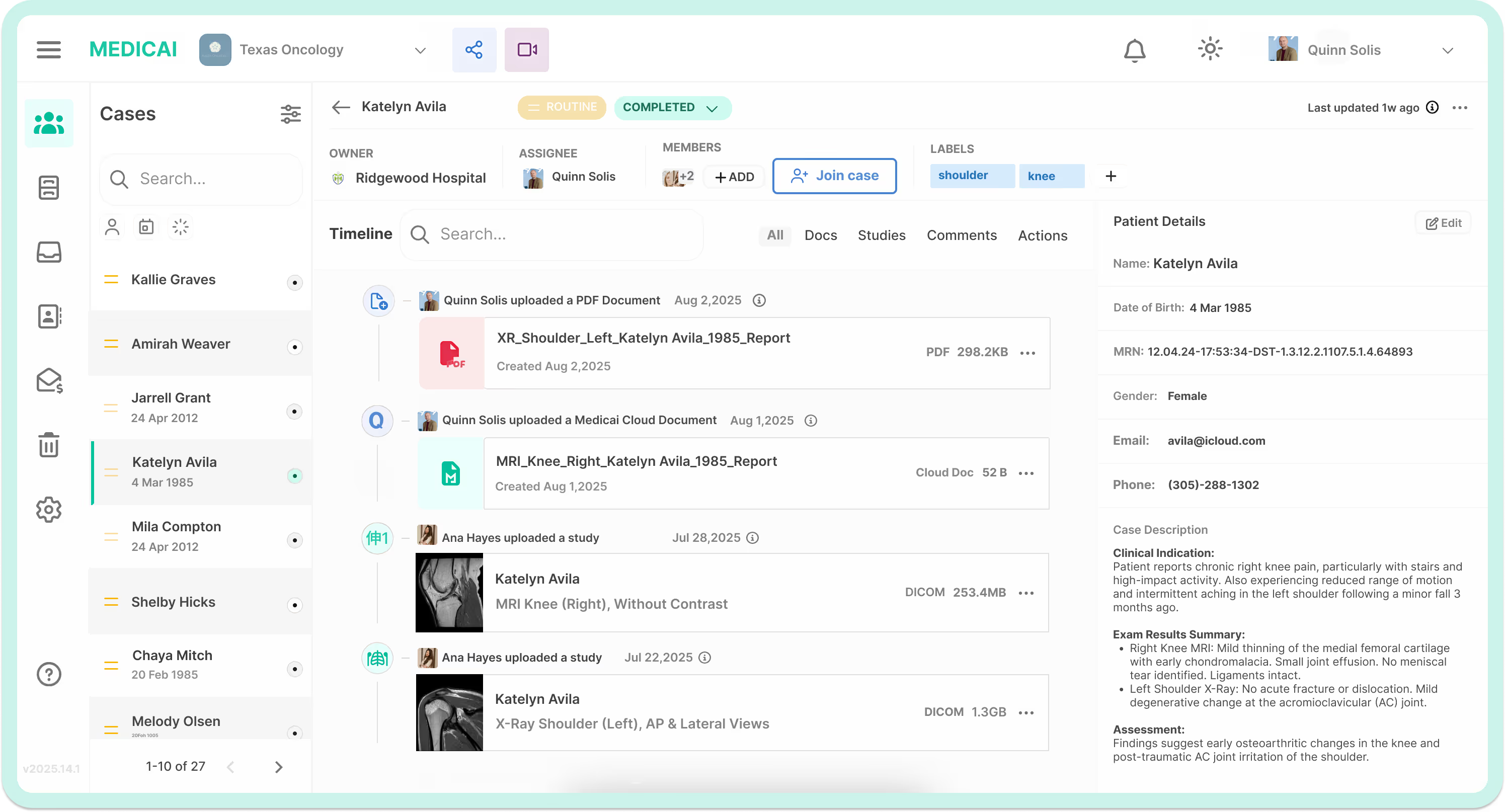
Experience unparalleled image clarity, advanced 3D reconstructions, and seamless integration with our scalable Cloud PACS, web and mobile DICOM viewer. Our solution streamlines neuroimaging workflows, optimizes collaboration, and enhances diagnostic accuracy, improving patient outcomes and operational efficiency. Elevate your workflow and deliver confident diagnoses.


Medicai's Neurology Solutions include tools and features to meet the unique needs of neurologists. These include Cloud PACS, DICOM Viewer, Neurology Patient Portal, Imaging Exchange, Medical Imaging Uploader. Our cloud based solution makes analyze brain scans, pinpoint stroke locations, and track MS progression easy for the neuroimaging experts and collaborate instantly with colleagues across the globe.


A specialized platform giving secure access to their medical imaging reports and images and enhancing communication with their doctors.
Makes it easier for neurologists and other healthcare providers to share neuroimaging data, fostering effective communication and teamwork.
It makes it easy for doctors to retrieve patient records by providing a flexible and safe uploading option for neuroimaging data.
Includes features that streamline workflows and save neurology specialists' time, such as the ability to view investigations instantly and share them online.
Medicai's integrated PACS streamlines neuroimaging workflow, enhances decision-making, and ultimately improves patient outcomes. Experience seamless access to critical brain images, collaborate instantly with specialists, and leverage AI-powered tools for faster, more accurate diagnoses.
PACS eliminates the need for physical film storage, allowing for rapid access to medical images from any location within the healthcare facility. This quick retrieval speeds up the diagnostic process and treatment planning.
With high-definition images available for review, PACS allows healthcare providers to zoom and pan through images, providing a more comprehensive view. This capability leads to more accurate diagnoses.
PACS facilitates improved communication among healthcare providers by enabling sharing of images across departments. This collaboration is essential in neurology, where interdisciplinary teams often need to work together to develop effective treatment plans.
While the initial investment in PACS can be significant, the long-term benefits include reduced costs associated with physical storage and improved diagnostic efficiency. By minimizing the need for repeat imaging and enhancing workflow, PACS can lower overall healthcare expenses.
PACS ensures secure long-term storage of medical images, reducing the risk of data loss and ensuring compliance with health data regulations. This secure storage is vital for maintaining patient history and facilitating ongoing care.
Choosing Medicai's Neurology PACS Solutions means embracing a comprehensive and tailored approach to managing neuroimaging data. Medicai empowers neurology doctors to optimize their workflow, enhance collaboration with other healthcare providers, and ensure the highest quality of patient care.
Now doctors can efficiently handle neuroimaging data with Medicai's Neurology Solutions, which operate within a user-friendly cloud platform with intuitive tools and features.
Neurology physicians can quickly interact with other medical professionals, retrieve and examine neuroimaging studies, and send investigations online to other parties thanks to advanced automation and seamless connectivity.
Our multi-enterprise solution enables modern neurology practices to automatically retrieve imaging from their own PACS and modalities, connect to partners, and allow their patients to easily upload their previous imaging studies.

Medicai's interoperable imaging infrastructure securely and compliantly store all the studies, along with complementary files such as reports, images, and videos, adhering to HIPAA and GDPR regulations. Thanks to our robust API and our granular access control level, the data is readily available. This makes the implementation of advanced neurology imaging workflows a breeze.

Team members can access, view, and collaborate easily with our ready-made web portal for doctors and mobile apps for doctors. They can also share their cases with specialists outside the organization in a secure and compliant manner. The Patient Portal enables your clinic to share imaging data with its patients. Tech-savvy neurology practices leverage our APIs and our DICOM components to embed DICOM capabilities into their existing web portals and apps.

Give it a try, play with it! Using our embeddable DICOM Viewer, you can easily view your DICOM files anywhere online (web, in the mobile application). Your DICOM files are stored in your Medicai workspace, in your cloud PACS.


"Definitely this application provides us with fast, remote imaging acquisitions. In addition, we can always make comparisons between different cases, we can make databases, we can make collections on different pathologies, we can collect special cases, I mean it's actually a mini real-time library, extremely, extremely useful for neurologists, for neurosurgeons, for surgeons, for oncology, for hematology."
Armand Frăsineanu, MD

Medicai's PACS solution empowers neurologists with the efficiency, accuracy, and collaboration needed to deliver exceptional patient care. Experience the power of seamless image access, advanced visualization tools, and AI-driven diagnostics.


What are the latest neuroimaging techniques advancing neuroscience research?
Medicai is at the forefront of incorporating cutting-edge neuroimaging techniques that are shaping the future of neuroscience. This includes advanced modalities such as functional ultrasound imaging, which offers high-resolution images of brain activity, and axial tomography for detailed structural views of the brain. In psychiatry and cognitive neuroscience, bold imaging and functional connectivity analyses provide insights into brain network operations during various mental states.
Moreover, Medicai leverages techniques like myelography and pneumoencephalography, specialized procedures that improve the visualization of the central nervous system and assist in diagnosing disorders. Additionally, the Willis blood flow model is applied in studies using transcranial Doppler to assess cerebrovascular reactivity, helping researchers and clinicians understand cerebral blood flow dynamics in healthy controls and patients with cerebrovascular diseases.
These advancements are supported by robust data analysis tools that enhance the accuracy of diagnostics and research findings. Medicai remains committed to advancing medical specialty through continuous innovation and by following the latest research and developments published in leading neuroscience journals. Our commitment extends to collaborations with top institutions, including neuroscience at King, to push the boundaries of what's possible in medical imaging and patient care.
What is the Significance of Synthetic Brain Imaging in Neuroscience?
Synthetic brain imaging is a groundbreaking approach in cognitive neuroscience that utilizes artificial constructs to simulate brain imaging data. This method allows researchers to model complex brain functions, such as neuronal activity and neurovascular responses, providing a controlled environment to study cerebral functions and disorders.
How is Cerebral Blood Flow Measured in Clinical Settings?
Techniques like transcranial Doppler and cerebral vasomotor reactivity response are key in measuring cerebral blood flow in patients undergoing various treatments, including statin treatment intensity. These methods assess the dynamism of blood flow in the brain, important in both research settings and clinical assessments of cerebrovascular health.
What Role Does Neuroradiology Play in the Resection of Brain Tumors?
Neuroradiology, through the use of advanced brain imaging techniques such as structural MRI and functional MRI (fMRI), provides critical information that aids neurosurgeons in the precise resection of brain tumors. These imaging modalities offer detailed insights into tumor location, neurovascular coupling, and surrounding brain areas, essential for successful surgical outcomes.
How Does Functional Brain Imaging Support Patients with Alzheimer's?
Functional brain imaging, including methods like magnetic resonance imaging (MRI) and positron emission tomography (PET), plays a crucial role in diagnosing and monitoring Alzheimer's disease. These techniques enable the detection of pathological contrast enhancement and biomarkers that indicate the progression of Alzheimer's, thereby aiding in personalized medicine approaches for these patients.
What Are the Latest Advances in Neuroimaging and Their Applications in Decision Making?
Recent advances in neuroimaging, particularly in functional brain imaging techniques like fMRI and EEG, have significantly enhanced our understanding of neural activity and decision-making processes. These advancements allow neuroscientists and psychologists to explore the intricate link between brain imaging data and cognitive psychology, providing deeper insights into how decisions are made.
What is Functional Neuroimaging or functional Magnetic Resonance Imaging (fMRI)?
Functional neuroimaging plays a crucial role in both fundamental neuroscience and clinical neuroscience, bridging the gap between research innovation and practical medical applications. This branch of neuroscience uses techniques such as functional Magnetic Resonance Imaging (fMRI) and Magnetic Resonance Spectroscopy to observe and measure brain areas involved in various cognitive functions as well as assess brain health. These methods are instrumental in the study of brain development, neurological disorders, and diseases such as Alzheimer's.
Neuroscience departments at various institutions use functional neuroimaging not only for clinical imaging but also to improve neurosurgeons and clinicians' education and training. The history of neuroimaging is rich with advancements that have transformed neuroscience and neuroimaging into critical components of modern medicine, aiding in the diagnosis and management of conditions like ischemic stroke and cerebrovascular disease.
Articles in prominent journals often highlight how functional neuroanatomy and clinical research through neuroimaging tools are fundamental in developing treatment strategies. Moreover, this technology is vital for neuroscientists who conduct clinical significance assessments, particularly when evaluating patients with complex cerebrovascular diseases. As research continues to evolve, AI and research methods are increasingly applied within functional imaging studies, improving the precision and efficiency of diagnostic processes and patient resources management.
Is neuroimaging reliable evidence?
Neuroimaging can provide reliable evidence for diagnosing and understanding neurological conditions, but its interpretation can be complex and context-dependent. While neuroimaging offers valuable insights into brain structure and function, its findings must be integrated with other clinical data and expert analysis to make accurate medical or legal determinations.
Is neuroimaging harmful?
Neuroimaging techniques like MRI scanning and CT scans are generally safe when used appropriately. However, CT scans expose patients to a small amount of radiation, which carries a minimal risk. MRI involves no radiation but may be unsuitable for patients with certain implants or claustrophobia. It's important to discuss any risks with your healthcare provider.
Is EEG considered neuroimaging?
EEG, or Electroencephalography, is considered a form of neuroimaging that measures electrical activity in the brain. Unlike imaging techniques that visualize brain structure, the EEG focuses on brain function by recording electrical signals, providing valuable insights into brain activity patterns.
What are the 4 types of brain imaging?
The four main types of brain imaging are Magnetic Resonance Imaging (MRI), Computed Tomography (CT) scans, Positron Emission Tomography (PET) scans, and Electroencephalography (EEG). Each type provides different information about brain structure and function, aiding in the diagnosis and treatment of neurological conditions.
Seamlessly retrieve, view, store, and share medical imaging data with a robust multi-location, cloud PACS storage, zero-footprint DICOM viewers, AI support, and best-in-class sharing capabilities.